In the intricate metropolis of the human body, a constant, silent drama unfolds. Billions of cells work in concert, orchestrated by an unseen hand, maintaining a delicate balance vital for life. Among the most crucial elements in this biological symphony is calcium, a mineral often hailed as the cornerstone of strong bones and teeth. Yet, calcium is a mineral of paradox, a dual-natured entity capable of both sustaining life and subtly undermining it. It is here, at the crossroads of calcium’s destiny, that our story begins – the tale of a humble, often-overlooked micronutrient, Vitamin K, the ultimate traffic controller, directing this vital mineral away from peril and towards its rightful place.
Imagine for a moment a bustling city. Its lifeblood – the traffic – flows through a complex network of arteries and avenues. For optimal function, this traffic must be directed efficiently, keeping major thoroughfares clear and ensuring resources reach their intended destinations. Now, picture calcium as a crucial, multi-purpose cargo. It’s essential for construction, but if it veers off course, if it accumulates where it shouldn’t, it can cause devastating congestion, blockages, and structural damage. For too long, we’ve understood calcium’s role in building our skeletal infrastructure, but largely overlooked the sophisticated system that prevents it from becoming a menace elsewhere. The narrative of Vitamin K as a mere blood-clotting agent is a gross understatement, akin to describing a symphony conductor solely by their ability to keep time. It is a story of profound systemic regulation, a molecular maestro orchestrating calcium’s journey with precision and purpose.
Chapter 1: The Unseen Battle – Calcium’s Dual Nature
Calcium. The word immediately conjures images of milk, strong bones, and healthy smiles. And rightly so. Calcium is the most abundant mineral in the human body, forming the rigid matrix of our bones and teeth, providing structural integrity, and acting as a vast reservoir for systemic needs. Beyond its architectural role, calcium ions are indispensable intracellular messengers, facilitating muscle contraction, nerve signal transmission, hormone secretion, and countless enzymatic reactions. It is, unequivocally, a mineral of life.
Yet, this very essentiality harbors a dangerous duality. While bone requires calcium, our soft tissues, particularly the delicate and vital lining of our arteries, do not. The misdirection of calcium, its errant deposition in places it was never meant to be, represents one of the most insidious threats to human health. This phenomenon is broadly termed ectopic calcification, and its most clinically significant manifestation is vascular calcification.
Vascular calcification is not simply the accumulation of inert mineral deposits; it is an active, regulated process resembling bone formation, occurring within the walls of our blood vessels. It begins microscopically, with tiny specks of calcium phosphate, akin to nascent bone crystals, forming within the arterial wall. Over time, these microcalcifications can coalesce, grow, and harden, transforming pliable, elastic arteries into rigid, brittle tubes. This stiffening has profound consequences.
In the inner layer of the artery, the intima, calcification is a hallmark of atherosclerosis. Here, calcium intertwines with cholesterol plaques, contributing to their hardening and instability, making them more prone to rupture and subsequent clot formation, leading to heart attacks and strokes. In the middle layer, the media, calcification, often seen in conditions like chronic kidney disease and diabetes, leads to Mönckeberg’s arteriosclerosis. This form primarily causes arterial stiffening, increasing pulse wave velocity, raising systolic blood pressure, and placing a greater workload on the heart, ultimately contributing to heart failure and cardiovascular mortality.
The irony, the cruel twist in this tale, is that while calcium accumulates dangerously in our arteries, it is often simultaneously being leached from our bones. This is the calcium paradox: individuals with extensive arterial calcification frequently suffer from low bone mineral density and an increased risk of osteoporosis. Our body, it seems, is struggling to direct calcium effectively, allowing it to wreak havoc in one system while starving another. This paradox highlights a fundamental flaw in the prevailing wisdom that simply consuming more calcium, often with Vitamin D, is the sole solution for bone health. Without a proper director, the cargo is delivered to the wrong address, leading to both famine and flood.
Chapter 2: Enter the Maestro – Vitamin K and Its Forms
For decades, Vitamin K was largely relegated to a single, albeit critical, role: blood clotting. Its very name, derived from the German "Koagulationsvitamin," underscored this understanding. We knew that without Vitamin K, our blood would not clot, leading to uncontrolled bleeding. This function is primarily attributed to Vitamin K1, or phylloquinone, found abundantly in green leafy vegetables like spinach, kale, and broccoli. K1 acts in the liver, enabling the carboxylation of specific proteins necessary for coagulation. Its swift action and rapid turnover in the liver meant that research initially focused almost exclusively on this hepatic role.
But the story of Vitamin K, like any good epic, has a hidden chapter, a deeper truth that took longer to uncover. It turns out that Vitamin K is not a singular entity but a family of fat-soluble vitamins, united by a common chemical structure but differentiated by their side chains. While K1 commands the clotting cascade, another, more elusive branch of the family tree, Vitamin K2, or menaquinones (MK-n), was quietly orchestrating far more profound and widespread systemic effects, particularly in bone and cardiovascular health.
Vitamin K2 is a diverse group, characterized by a varying number of isoprene units in its side chain (MK-4, MK-7, MK-9, etc.). The most clinically significant forms are MK-4 and MK-7. MK-4 is a short-chain menaquinone, found in certain animal products like meat, eggs, and dairy, especially from grass-fed animals. It can also be synthesized endogenously from K1 in various tissues, though the efficiency of this conversion is debated. MK-7, a long-chain menaquinone, is predominantly found in fermented foods, most notably the Japanese soybean dish natto, which boasts extraordinarily high levels. Smaller amounts are found in other fermented products like certain cheeses.
The key distinction between K1 and K2, especially MK-7, lies in their pharmacokinetics and tissue distribution. K1, with its short half-life, is rapidly absorbed and primarily shunted to the liver for clotting factor activation. MK-7, however, has a much longer half-life, allowing it to circulate in the blood for extended periods and reach extra-hepatic tissues – bones, arteries, and other soft tissues – where it can exert its unique regulatory functions. This longer systemic availability is what makes MK-7 particularly effective as a "traffic controller" for calcium beyond the liver.
The recognition of K2’s broader role was a slow burn, a gradual accumulation of observational data and mechanistic insights that challenged the long-held dogma. Researchers began to notice that while K1 intake correlated with reduced risk of certain cancers, it showed little correlation with cardiovascular outcomes or bone density. K2, however, was a different story. Landmark studies, like the Rotterdam Study in the Netherlands, began to reveal a powerful association: higher dietary intake of K2 (specifically MK-7 and MK-9) correlated with significantly reduced arterial calcification, lower risk of cardiovascular disease, and improved overall survival, independent of K1 intake. These findings were nothing short of a paradigm shift, unveiling Vitamin K as a critical player in a metabolic dance far more complex than previously imagined. The maestro had finally stepped into the spotlight, ready to reveal its full repertoire.
Chapter 3: The Molecular Mechanism – Guiding the Traffic
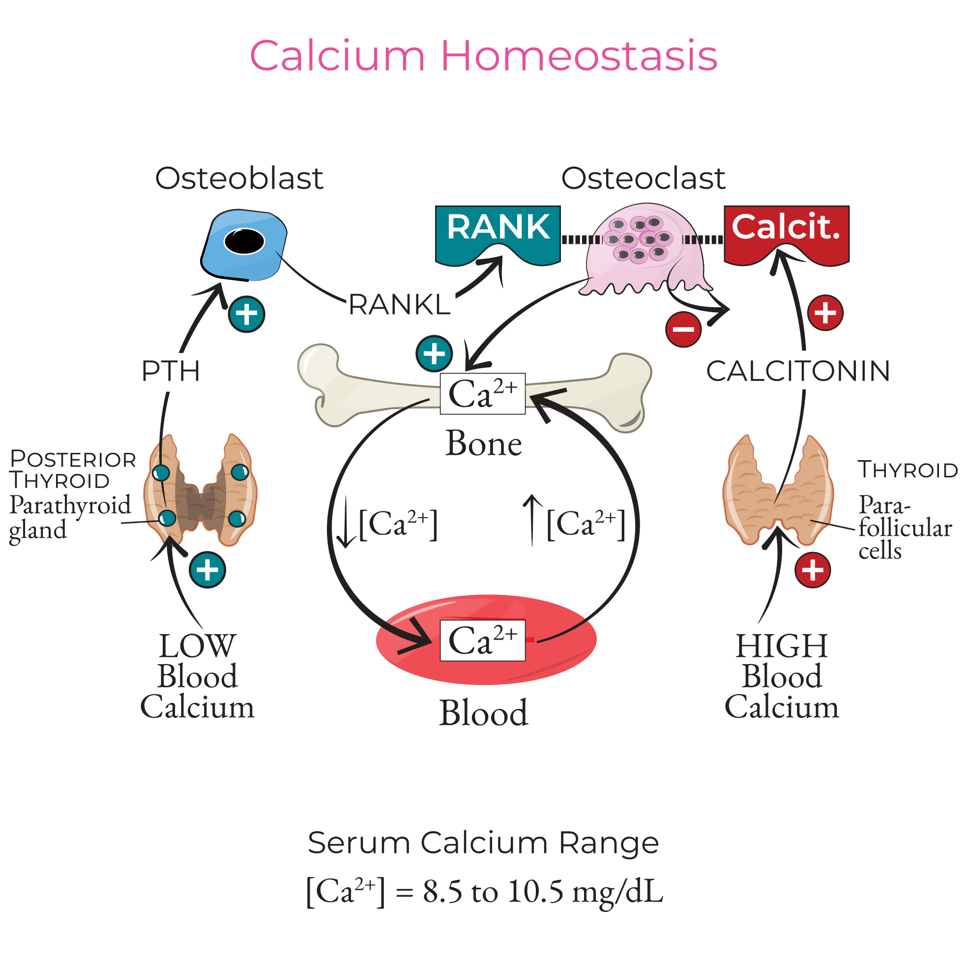
The genius of Vitamin K’s traffic control lies in its singular biochemical function: its role as an essential co-factor for an enzyme called gamma-glutamyl carboxylase (GGCX). This enzyme is responsible for a post-translational modification known as carboxylation. During this process, specific glutamic acid residues (a type of amino acid) on certain proteins are converted into gamma-carboxyglutamic acid (Gla) residues. These newly formed Gla residues possess a unique ability: they can bind calcium ions with high affinity. Without Vitamin K, GGCX cannot function, and these proteins remain in their uncarboxylated, inactive forms, unable to perform their calcium-binding tasks. These are the Gla proteins, and they are the unsung heroes, the molecular enforcers, that execute Vitamin K’s directives.
Among the pantheon of Gla proteins, two stand out as primary agents in the arterial-skeletal calcium traffic control system: Matrix Gla Protein (MGP) and Osteocalcin (BGP).
Matrix Gla Protein (MGP): The Arterial Guardian
Imagine MGP as the vigilant crossing guard of your arteries, ensuring no calcium cargo strays onto the vascular pathways. MGP is predominantly produced by vascular smooth muscle cells and chondrocytes (cartilage cells). Its primary function, when fully carboxylated by Vitamin K, is to inhibit calcification in soft tissues, especially the arteries.
How does it work? Carboxylated MGP acts as a potent local inhibitor of hydroxyapatite crystal formation. It binds tightly to calcium ions and nascent calcium crystals, effectively sequestering them and preventing their growth and aggregation within the arterial wall. Think of it as a molecular net, catching rogue calcium particles before they can embed themselves and form plaques. It’s a guardian, actively preventing the misdirection of calcium.
The critical implication here is that if there is insufficient Vitamin K (particularly K2) to carboxylate MGP, the protein remains in its inactive, uncarboxylated form (ucMGP). This "sleeping" guardian cannot bind calcium, and the arterial traffic is left unregulated. Calcium is then free to deposit in the arterial walls, initiating and propagating vascular calcification. Levels of ucMGP in the bloodstream are increasingly recognized as a biomarker for Vitamin K deficiency and a strong predictor of arterial stiffness and cardiovascular risk. When the MGP crossing guard is asleep, calcium traffic spirals out of control.
Osteocalcin (BGP): The Bone Builder
Now, let’s shift our gaze to the bone, the intended destination for calcium. Here, our second major Gla protein, Osteocalcin, plays a pivotal role. Produced by osteoblasts, the bone-building cells, osteocalcin is the chief non-collagenous protein in bone matrix. Its function, when fully carboxylated by Vitamin K, is to promote the integration of calcium into the bone structure.
Carboxylated osteocalcin is like the foreman at a construction site, directing the calcium cargo to its proper place within the bone matrix. It has a high affinity for hydroxyapatite, the mineral component of bone, and helps to orchestrate the crystallization and mineralization processes that lead to strong, dense bone. It essentially "locks" calcium into the bone.
Conversely, if Vitamin K is insufficient, osteocalcin remains largely uncarboxylated (ucOC). This inactive foreman cannot effectively guide calcium into the bone matrix. Consequently, calcium is not properly utilized for bone formation, potentially contributing to lower bone mineral density and an increased risk of osteoporosis. High levels of ucOC are therefore considered a marker of Vitamin K deficiency and a predictor of fracture risk. When the osteocalcin foreman is inactive, the bone construction site suffers from inefficient calcium delivery.
In essence, Vitamin K acts as the ultimate "on/off" switch for these vital proteins. By ensuring MGP is active, it removes calcium from arteries. By ensuring osteocalcin is active, it places calcium into bones. This dual action is the elegant solution to the calcium paradox, providing a biochemical pathway to both prevent arterial calcification and promote bone mineralization. It is the molecular manifestation of the traffic controller at its finest, intelligently directing calcium flow, preventing catastrophe in one area while fostering growth in another.
Other Gla proteins exist, such as Protein S (involved in both coagulation and bone metabolism) and Gas6 (involved in cell growth and adhesion), hinting at the broader, still-unfolding story of Vitamin K’s influence across various physiological systems. But for the purpose of calcium traffic control, MGP and Osteocalcin are the undisputed stars.
Chapter 4: The Traffic Controller in Action – Clinical Evidence and Impact
The molecular mechanisms, while elegant, gain true significance when backed by robust clinical evidence. The journey from biochemical discovery to real-world impact has been a long one for Vitamin K, but compelling data now firmly establishes its role as a crucial traffic controller.